Brain tissue of donors of the NBB-PSY program is collected by the NBB during autopsy. This is referred to as 'postmortem brain tissue'.

Lot contributed to the initiation of the Netherlands Brain Bank for Psychiatry (NBB-PSY). NBB-PSY is a brain donor and autopsy program of the Netherlands Brain Bank launched with the mission to increase the availability of tissue for fundamental and applied research into psychiatric disorders.


Microglia are isolated from postmortem brain tissue by creating a single cell suspension, followed by density gradient centrifugation, and further isolation of the cells using antibodies.
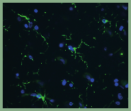
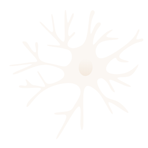
Microglia in brain tissue: Changes in microglial density, morphology and phenotype are assessed by staining postmortem brain tissue with markers for microglial cells
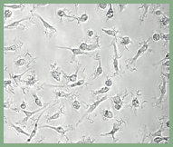
Isolated Microglia: After isolation, microglia are cultured in the lab to analyze their different functions.
Brain tissue of donors of the NBB-PSY program is collected by the NBB during autopsy. This is referred to as 'postmortem brain tissue'.
Lot contributed to the initiation of the Netherlands Brain Bank for Psychiatry (NBB-PSY). NBB-PSY is a brain donor and autopsy program of the Netherlands Brain Bank launched with the mission to increase the availability of tissue for fundamental and applied research into psychiatric disorders.
Microglia are isolated from postmortem brain tissue by creating a single cell suspension, followed by density gradient centrifugation, and further isolation of the cells using antibodies.
Microglia in brain tissue: Changes in microglial density, morphology and phenotype are assessed by staining postmortem brain tissue with markers for microglial cells
Isolated Microglia: After isolation, microglia are cultured in the lab to analyze their different functions.
Contribution of Age, Brain Region, Mood Disorder Pathology, and Interindividual Factors on the Methylome of Human Microglia.
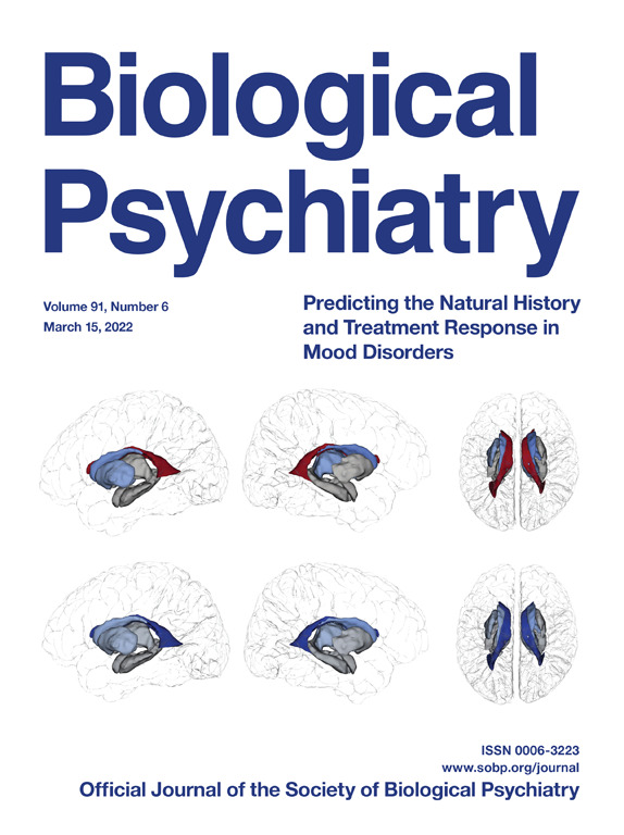
de Witte LD, Wang Z, Snijders GLJL, Mendelev N, Liu Q, Sneeboer MAM, Boks MPM, Ge Y, Haghighi F. Biol Psychiatry. 2022 Mar 15;91(6):572-581.
Genetic analysis of the human microglial transcriptome across brain regions, aging and disease pathologies.
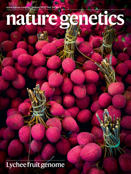
Lopes KP, Snijders GJL, Humphrey J, Allan A, Sneeboer MAM, Navarro E, Schilder BM, Vialle RA, Parks M, Missall R, van Zuiden W, Gigase FAJ, Kübler R, van Berlekom AB, Hicks EM, Bӧttcher C, Priller J, Kahn RS, De Witte LD*, Raj T*. Nature Genetics 2022 Jan;54(1):4-17. *Shared senior authors.
Single-cell mass cytometry of microglia in major depressive disorder reveals a non-inflammatory phenotype with increased homeostatic marker expression.
Böttcher C, Fernández-Zapata C, Snijders GJL, Schlickeiser S, Sneeboer MAM, Kunkel D, De Witte LD, Priller J. Transl Psychiatry. 2020 Sep 11;10(1):310.
Distinct non-inflammatory signature of microglia in post-mortem brain tissue of patients with major depressive disorder.
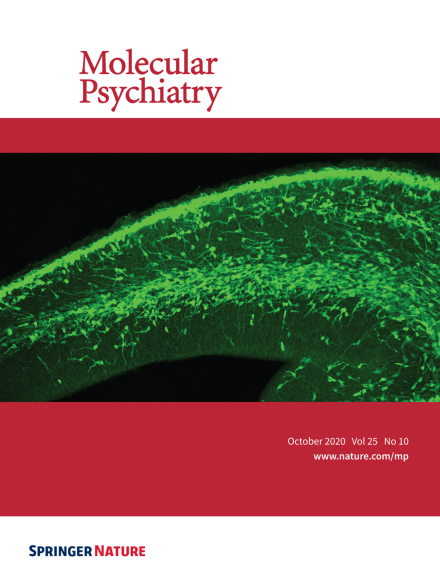
Snijders GJLJ, Sneeboer MAM, Fernández-Andreu A, Udine E; Psychiatric donor program of the Netherlands Brain Bank (NBB-Psy), Boks MP, Ormel PR, van Berlekom AB, van Mierlo HC, Bӧttcher C, Priller J, Raj T, Hol EM, Kahn RS, De Witte LD. Molecular Psychiatry. 2020 Oct 7
Microglial activation in schizophrenia: Is translocator 18 kDa protein (TSPO) the right marker?
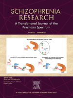
Sneeboer MAM, van der Doef T, Litjens M, Psy NBB, Melief J, Hol EM, Kahn RS, de Witte LD. Schizophr Res. 2019 Nov 4. pii: S0920-9964(19)30480-3. doi: 10.1016/j.schres.2019.10.045.
Increased number of T-lymphocytes in post-mortem brain tissue of patients with schizophrenia.
Sneeboer MAM, van Mierlo HC, Stotijn E, MacIntyre DJ, Smith C, Kahn RS, Hol EM, de Witte LD. Schizophr Res. 2019 Oct 29.
Microglia in post-mortem brain tissue of patients with bipolar disorder are not immune activated.
Sneeboer M, Snijders G, Berdowski W, Fernández-Andreu A, NBB- PSY, Van Mierlo H, Berdenis van Berlekom A, Litjens M, Kahn R, Hol E, and De Witte LD. Transl Psychiatry. 2019 May 24;9(1):153.
Deep immune profiling of human brain microglia by multiplexed mass cytometry.
Bottcher C, Schlickeiser S, Sneeboer M, Kunkel D, Knop A, Paza E, Fidzinski P, Krauss L, Snijders G, Kahn R, Schulz A, Mei H, NBB-PSY, Hol E, Siegmund B, Glauben R, Spruth E, de Witte L, Priller J..Nature Neuroscience, 2019 Jan;22(1):78-90. doi: 10.1038/s41593-018-0290-2
Characterizing primary human microglia: A comparative study with myeloid subsets and culture models.
Melief J, Sneeboer MA, Litjens M, Ormel PR, Palmen SJ, Huitinga I, Kahn RS, Hol EM, de Witte LD. Glia 2016 Jul 21. PMID: 27442614 DOI: 10.1002/glia.23023
The translation of genetic risk variants to molecular disease mechanisms.
Snijders G, Snijders R, de Witte LD. Tijdschr Psychiatr. 2022;64(5):323-326.