

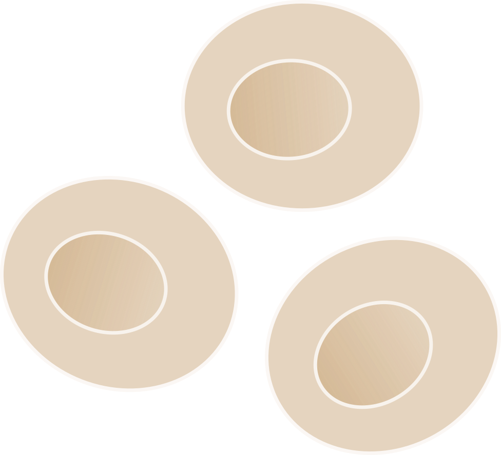
Patient-derived material can be reprogrammed to induced pluripotent stem cells, in short iPSCs. This discovery by Gurdon & Yamanaka was awarded with the nobel prize in physiology or medicine in 2012 and has since started a new research era of human stem cell disease modelling. We utilize this technology in our lab to understand the role of the immune system in brain diseases.
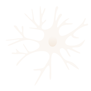
The resident immune cells in the central nervous system are called microglia. They are among the most versatile cells of the body and their high levels of plasticity allows for rapid responses to changes in the brain environment. Microglia are essential for brain function and therefore associated with a variety of neurological and psychiatric disorders
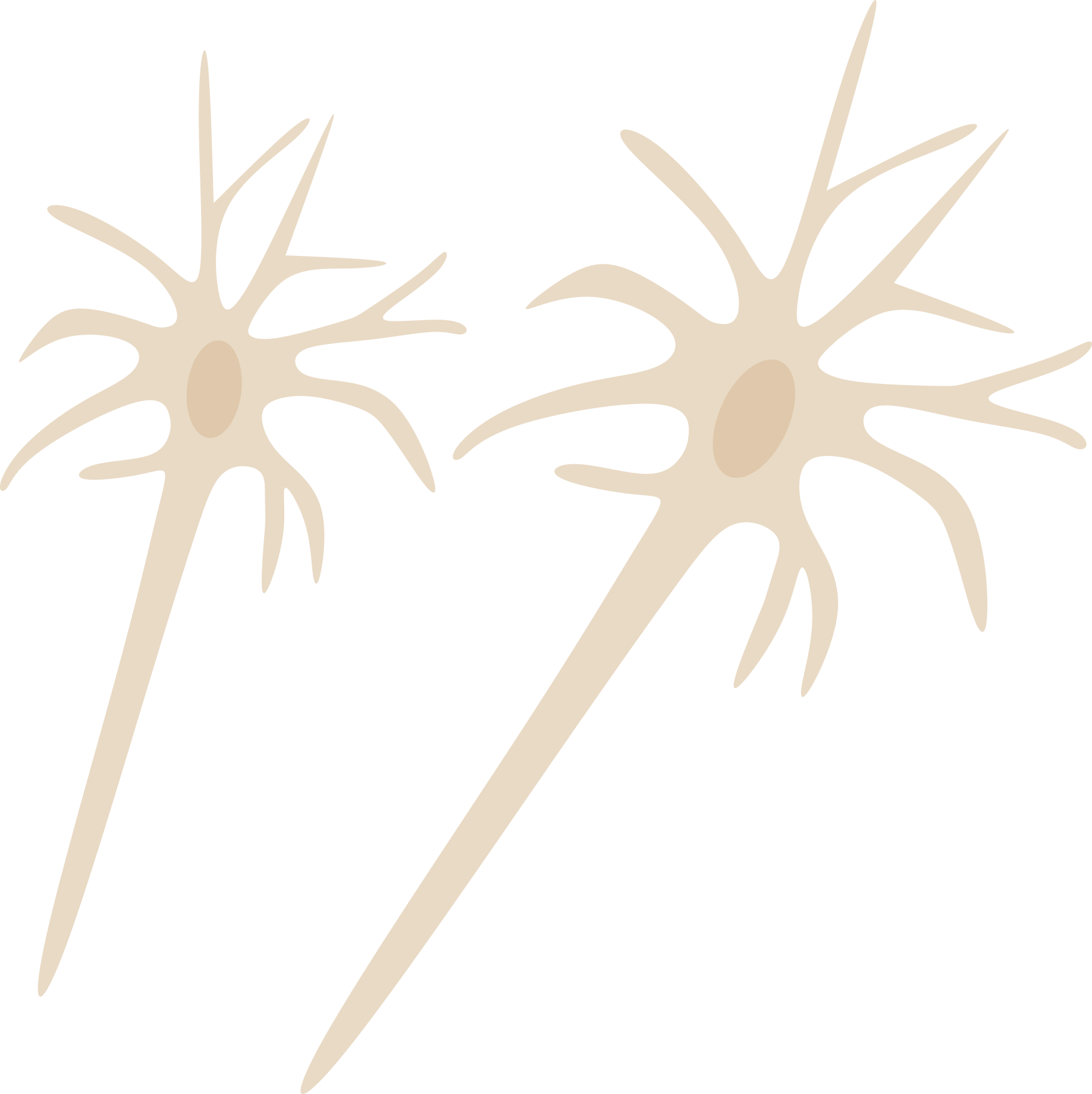
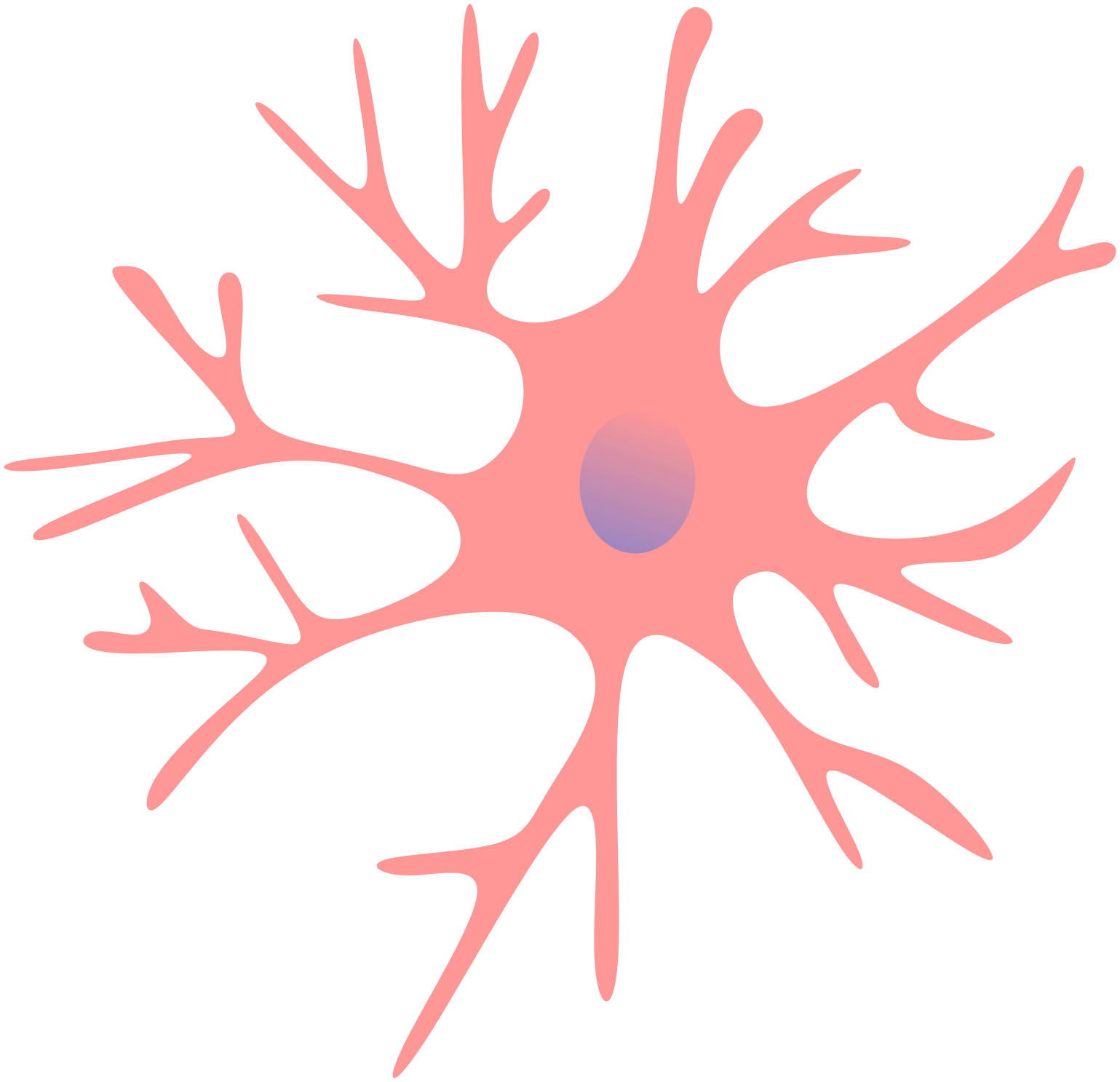
Neuronal cells are important for information processing in the brain and act in a complex interplay with microglia. We can model neuronal network development in our lab by using iPSCs and study their interaction with the immune system.
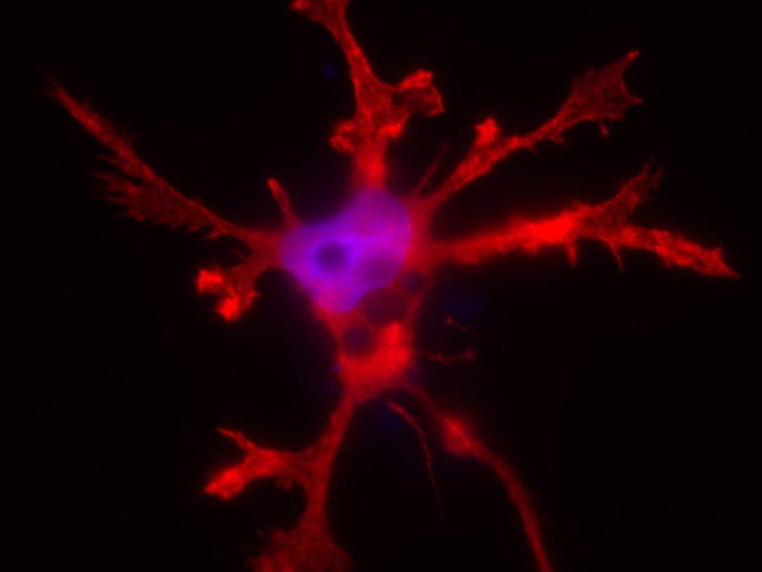
We use different techniques to study our stem cell models, one of them is fluorescent (live) imaging. Here, you can see how the microglia and neurons look like in our lab. Microglia show their characteristic ramifications allowing them to continuously scan their environment. The neurons formed a dense network with numerous connections, called synapses.
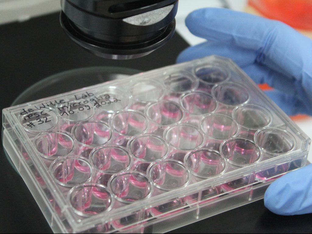
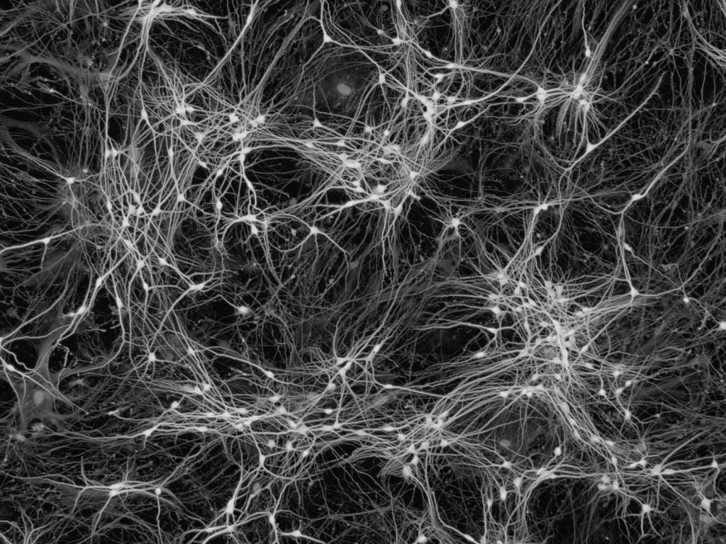
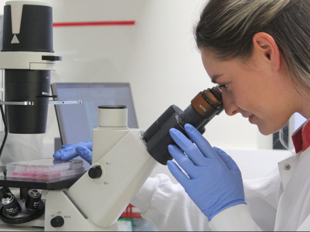
We use different techniques to study our stem cell models, one of them is fluorescent (live) imaging. Here, you can see how the microglia and neurons look like in our lab. Microglia show their characteristic ramifications allowing them to continuously scan their environment. The neurons formed a dense network with numerous connections, called synapses.
Exposure to the Amino Acids Histidine, Lysine, and Threonine Reduces mTOR Activity and Affects Neurodevelopment in a Human Cerebral Organoid Model.
Berdenis van Berlekom A, Kübler R, Hoogeboom JW, Vonk D, Sluijs JA, Pasterkamp RJ, Middeldorp J, Kraneveld AD, Garssen J, Kahn RS, Hol EM, de Witte LD, Boks MP. Nutrients. 2022 May 23;14(10):2175
Characterization of HIV-1 Infection in Microglia-Containing Human Cerebral Organoids.
Gumbs SBH, Berdenis van Berlekom A, Kübler R, Schipper PJ, Gharu L, Boks MP, Ormel PR, Wensing AMJ, de Witte LD, Nijhuis M. Viruses. 2022 Apr 16;14(4):829.
Human microglial models to study HIV infection and neuropathogenesis: a literature overview and comparative analyses.
Gumbs SBH, Kübler R, Gharu L, Schipper PJ, Borst AL, Snijders GJLJ, Ormel PR, van Berlekom AB, Wensing AMJ, de Witte LD, Nijhuis M. J Neurovirol. 2022 Feb;28(1):64-91.

Microglia innately develop within cerebral organoids.
Ormel PR, Vieira de Sá R, van Bodegraven EJ, Karst H, Harschnitz O, Sneeboer MAM, Johansen LE, van Dijk RE, Scheefhals N, Berdenis van Berlekom A, Ribes Martínez E, Kling S, MacGillavry HD, van den Berg LH, Kahn RS, Hol EM*, de Witte LD*, Pasterkamp RJ*, Nature Communications, 2018 Oct 9;9(1):4167. *Senior authors.
A characterization of the molecular phenotype and inflammatory response of schizophrenia patient-derived microglia-like cells.
Ormel PR, Böttcher C, Gigase FAJ, Missall RD, van Zuiden W, Fernández Zapata MC, Ilhan D, de Goeij M, Udine E, Sommer IEC, Priller J, Raj T, Kahn RS, Hol EM, de Witte LD. Brain Behav Immun. 2020 Nov;90:196-207. doi: 10.1016/j.bbi.2020.08.012. Epub 2020 Aug 13.